|
Instructions for Huida Glue-Free Quick-Drying Microscope Cover Glass
I. Product Overview
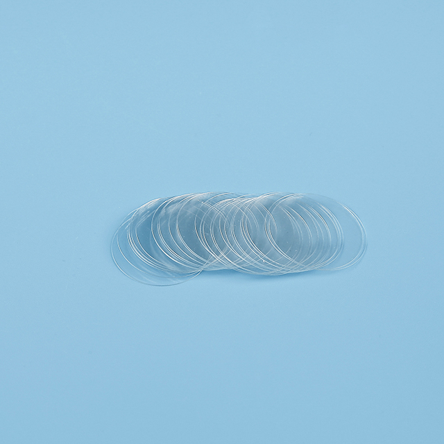
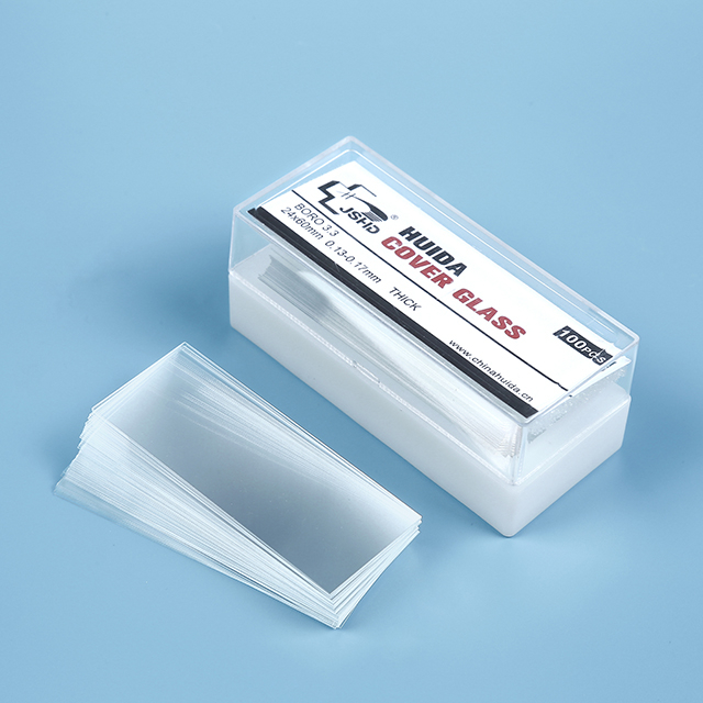
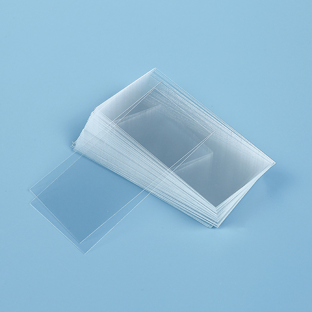
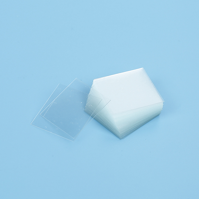
The Huida Glue-Free Quick-Drying Microscope Cover Glass are specially designed for efficient and glue-free fixation. They achieve rapid adhesion through physical coating technology, suitable for pathological, cytological, and molecular biology experiments. Their core advantages include no need for glue, fast operation, and compatibility with automated equipment.
II. Technical Features
1. Glue-Free Fixation Technology: Fixed via physical coating, eliminating the need for glue.
2. High-Precision Manufacturing: Compatible with standard microscope objectives, such as oil immersion lens.
3. Flatness: Surface flatness tolerence ≤ ± 1 μm to avoid imaging distortion.
4. Anti-Detachment Coating: Suitable for long-term preserved samples such as paraffin sections.
5. Compatibility: Supports precise robotic arm picking and fits most microscope slides.
III. Usage Steps
1. Sample Preparation
- Liquid samples (e.g., cell suspensions, blood): Add 1~2 drops (50~100 μL) to the center of the slide.
- Solid samples (tissue sections): Gently press to fix and ensure flatness.
2. Placing the Cover Glass
- Tilting Method (Universal): Hold the edge of the cover slip with forceps, touch one end to the liquid drop first, then slowly tilt and lower it to spread the liquid via surface tension.
Note: Avoid touching the coated area with fingers.
- Electrostatic Adsorption Method (for some models): Gently touch the cover glass to the slide and fix it via electrostatic adsorption without pressing.
3. Bubble Handling
- Small bubbles can dissipate naturally or be expelled by gently pressing the edges with forceps.
- Large bubbles require repositioning the cover glass.
4. Drying and Fixation
- Natural drying: Let stand for 5-6 minutes; the liquid fixes via surface tension.
- Accelerated drying (optional): Assist with low-wind hot air (≤37°C) to avoid sample damage from high temperatures.
5. Subsequent Operations
- Staining/Sealing: Directly drop stain or mounting medium (e.g., xylene, glycerin) to cover the area of the cover slip.
- Microscope observation: Ensure the cover glass is secure; gently press the slide under high-power lens to prevent displacement.
IV. Application Scenarios
1. Pathological Diagnosis
- Rapid sealing of tissue sections (HE staining, immunohistochemistry), with anti-detachment coating reducing the risk of section loss.
2. Molecular Biology
- FISH or ISH experiments, where the hydrophobic coating prevents probe diffusion.
3. Automated Platforms
- Compatible with fully automated equipment such as Ventana, suitable for precise robotic arm operations.
V. Precautions
1. Store long-term in a dry, light-proof environment to prevent coating aging.
2. For the hydrophobic coating: Avoid cleaning with strong acids or alkalis (may damage the coating).
VI. Common Problem Solutions
Questions | Solutions |
Cover Glass sliding or displacement | Check whether the coating is worn and ensure the correct operation of the electrostatic adsorption model |
Sample overflow or uneven distribution | Reduce the dropping volume to 30-50 μL, and control the tilting angle at 30°~45° |
Coating peeling or scratching | Stopping using and replace a new cover slip |
Bubbles are difficult to expel | Gently squeeze for the edge with forceps to avoid direct puncture |
VII. Model Selection Recommendations
- Routine HE Staining: Choose the standard 0.17 mm thickness hydrophobic coating model.
- Immunohistochemistry (IHC): Select the APES anti-detachment coating type, suitable for high-temperature baking.
- Fluorescence Experiments: Use the fluorescence anti-quenching coating model to avoid organic mounting media.
- Automated Platforms: Confirm the equipment model and choose the Huida-Ventana compatible model.
Huida Glue-Free Quick-Drying Cover Glass, with their high precision, fast operation, and wide compatibility, are ideal for pathological, research, and clinical diagnostic applications. For specific model parameters, please refer to the official instruction manual or contact the supplier for technical support.
Instructions for Huida Glue-Free Quick-Drying Microscope Cover Glass
I. Product Overview
The Huida Glue-Free Quick-Drying Microscope Cover Glass are specially designed for efficient and glue-free fixation. They achieve rapid adhesion through physical coating technology, suitable for pathological, cytological, and molecular biology experiments. Their core advantages include no need for glue, fast operation, and compatibility with automated equipment.
II. Technical Features
1. Glue-Free Fixation Technology: Fixed via physical coating, eliminating the need for glue.
2. High-Precision Manufacturing: Compatible with standard microscope objectives, such as oil immersion lens.
3. Flatness: Surface flatness tolerence ≤ ± 1 μm to avoid imaging distortion.
4. Anti-Detachment Coating: Suitable for long-term preserved samples such as paraffin sections.
5. Compatibility: Supports precise robotic arm picking and fits most microscope slides.
III. Usage Steps
1. Sample Preparation
- Liquid samples (e.g., cell suspensions, blood): Add 1~2 drops (50~100 μL) to the center of the slide.
- Solid samples (tissue sections): Gently press to fix and ensure flatness.
2. Placing the Cover Glass
- Tilting Method (Universal): Hold the edge of the cover slip with forceps, touch one end to the liquid drop first, then slowly tilt and lower it to spread the liquid via surface tension.
Note: Avoid touching the coated area with fingers.
- Electrostatic Adsorption Method (for some models): Gently touch the cover glass to the slide and fix it via electrostatic adsorption without pressing.
3. Bubble Handling
- Small bubbles can dissipate naturally or be expelled by gently pressing the edges with forceps.
- Large bubbles require repositioning the cover glass.
4. Drying and Fixation
- Natural drying: Let stand for 5-6 minutes; the liquid fixes via surface tension.
- Accelerated drying (optional): Assist with low-wind hot air (≤37°C) to avoid sample damage from high temperatures.
5. Subsequent Operations
- Staining/Sealing: Directly drop stain or mounting medium (e.g., xylene, glycerin) to cover the area of the cover slip.
- Microscope observation: Ensure the cover glass is secure; gently press the slide under high-power lens to prevent displacement.
IV. Application Scenarios
1. Pathological Diagnosis
- Rapid sealing of tissue sections (HE staining, immunohistochemistry), with anti-detachment coating reducing the risk of section loss.
2. Molecular Biology
- FISH or ISH experiments, where the hydrophobic coating prevents probe diffusion.
3. Automated Platforms
- Compatible with fully automated equipment such as Ventana, suitable for precise robotic arm operations.
V. Precautions
1. Store long-term in a dry, light-proof environment to prevent coating aging.
2. For the hydrophobic coating: Avoid cleaning with strong acids or alkalis (may damage the coating).
VI. Common Problem Solutions
Questions | Solutions |
Cover Glass sliding or displacement | Check whether the coating is worn and ensure the correct operation of the electrostatic adsorption model |
Sample overflow or uneven distribution | Reduce the dropping volume to 30-50 μL, and control the tilting angle at 30°~45° |
Coating peeling or scratching | Stopping using and replace a new cover slip |
Bubbles are difficult to expel | Gently squeeze for the edge with forceps to avoid direct puncture |
VII. Model Selection Recommendations
- Routine HE Staining: Choose the standard 0.17 mm thickness hydrophobic coating model.
- Immunohistochemistry (IHC): Select the APES anti-detachment coating type, suitable for high-temperature baking.
- Fluorescence Experiments: Use the fluorescence anti-quenching coating model to avoid organic mounting media.
- Automated Platforms: Confirm the equipment model and choose the Huida-Ventana compatible model.
Huida Glue-Free Quick-Drying Cover Glass, with their high precision, fast operation, and wide compatibility, are ideal for pathological, research, and clinical diagnostic applications. For specific model parameters, please refer to the official instruction manual or contact the supplier for technical support.